Chuyển tới nội dung
Trang chủ » Fat Pad Sign: A Clue To Pericardial Effusion

Fat Pad Sign: A Clue To Pericardial Effusion

Pericardial Effusion | Radiology Reference Article | Radiopaedia.Org

What is the pericardial fat pad sign?

The pericardial fat pad sign is a nice but rare sign of pneumothorax on supine CXR. The pericardial fat displaces laterally as the lung is no longer adjacent to it pushing it against the mediastinum, hence causing a lumpy appearance of the cardiac borders.

What is the difference between pericardial effusion and pericardial fat pad?

Echocardiographically, pericardial fat is a noncircumferential accumulation of ultrasonographically heterogeneous material that moves in concert with the heart. In contrast, pericardial effusions are typically stationary, echolucent, and circumferential rather than restricted to the region around the right heart.

What sign is seen in pericardial effusion?

If pericardial effusion signs and symptoms do occur, they might include: Shortness of breath or difficulty breathing (dyspnea) Discomfort when breathing while lying down. Chest pain, usually behind the breastbone or on the left side of the chest.

What is the sandwich sign of pericardial effusion?

Pericardial fat stripe, also known as sandwich or oreo cookie sign: The epicardial (red arrows) and paracardial fat (blue arrows) is separated by a “stripe” of increased density (light green *), representing pericardial fluid.

What is the fat pad sign and sail sign?

The fat pad sign, also known as the sail sign, is a potential finding on elbow radiography which suggests a fracture of one or more bones at the elbow. It is may indicate an occult fracture that is not directly visible. Its name derives from the fact that it has the shape of a spinnaker (sail).

What is the positive anterior fat pad sign?

The sail sign on an elbow radiograph, also known as the anterior fat pad sign, describes the elevation of the anterior fat pad to create a silhouette similar to a billowing spinnaker sail from a boat. It indicates the presence of an elbow joint effusion.

What is the triad of pericardial effusion?

Classic teaching is that patients with pericardial effusion will have muffled heart sounds and that Beck’s triad (hypotension, distended neck veins and muffled heart sounds) will be present in patients with pericardial tamponade.

What is Dressler syndrome?

Dressler syndrome is inflammation of the sac surrounding the heart (pericarditis). It’s believed to occur as the result of the immune system responding to damage to heart tissue or damage to the sac around the heart (pericardium). The damage can result from a heart attack, surgery or traumatic injury.

What is the hallmark of pericardial effusion?

The hallmark of effusive-constrictive pericarditis is the persistence of elevated right atrial pressure after removal of the pericardial fluid.

What is the Oreo cookie sign pericardial effusion?

A lateral film may show the Oreo cookie sign. This refers to the combination of fluid between the epicardial (anterior) and pericardial fat (posterior), resembling an Oreo cookie.

What are the criteria for pericardial effusion?

The main symptoms of pericardial effusions and cardiac tamponade include: Shortness of breath (dyspnea). Chest pressure or pain. Fast heartbeat or heart palpitations.

What is the Ewart sign in pericardial effusion?

In patients with large pericardial effusions, the Ewart sign may be present. This is an area of dullness with bronchial breath sounds heard just below the left scapula. The JVP tracing may reveal an absent ‘y’ descent due to the elevated intrapericardial pressure that prevents the filling of the ventricles.

What is the water bottle sign in pericardial effusion?

The water bottle sign or configuration refers to the shape of the cardiac silhouette on erect frontal chest x-rays in patients who have a very large pericardial effusion. Typically the effusion has accumulated over many weeks to months (e.g. in patients with malignancy) and the pericardium has gradually stretched.

What is the hallmark sign of pericarditis?

If you have pericarditis, the most common symptom is chest pain. This chest pain may: feel sharp or stabbing (however some people have dull, pressure-like chest-pain) be felt on the left-hand side of the chest or behind your breastbone.

What is the fat pad sign?

The posterior fat pad sign is the visualization of a lucent crescent of fat located in the olecranon fossa on a true lateral view of an elbow joint with the elbow flexed at a right angle indicating an elbow joint effusion1.

What is the cardiac fat pad sign?

The floating cardiac fat pad sign occurs when pleural air collects anteriorly and superiorly in the most non-dependent portion of the chest lifting the pericardial fat pad off the diaphragm. Lung markings are still seen surrounding the pericardial fat pad due to the inflated lower lobe of the lung resting dependently.

What is the PQ fat pad sign?

The pronator quadratus sign, also known as MacEwan sign, can be an indirect sign of distal forearm trauma. It relies on displacement of the fat pad that lies superficial to the pronator quadratus muscle as seen on a lateral wrist radiograph.

What is the fat pad separation sign?

Fat pad separation sign refers to the separation of the anterior suprapatellar and posterior suprapatellar (prefemoral) fat pads on lateral knee radiograph.

What is a fat pad?

A fat pad (aka haversian gland) is a mass of closely packed fat cells surrounded by fibrous tissue septa. They may be extensively supplied with capillaries and nerve endings. Examples are: Intraarticular fat pads. These are also covered by a layer of synovial cells.

What is Hoffa’s fat pad impingement sign?

Patients with Hoffa pad impingement syndrome report a “burning” or “aching” sensation to the anterior knee localized deep to and on either side of the patellar tendon adjacent to the inferior pole of the patella. This most commonly occurs with the knee at full extension, dynamic extension, or prolonged flexion.

How to check for pericardial effusion?

Numerous imaging techniques are utilized to evaluate pericardial effusion (PE) including chest X-ray, electrocardiogram, transthoracic echocardiography, CT scan, cardiac MRI, and pericardiocentesis.

How is pericardial effusion confirmed?

To diagnose pericardial effusion, the health care provider will typically perform a physical exam and ask questions about your symptoms and medical history. He or she will likely listen to your heart with a stethoscope. If your health care provider thinks you have pericardial effusion, tests can help identify a cause.

What are the signs of pericardial effusion on auscultation?

Patients with pericardial effusion may have unremarkable physical exams but often present with tachycardia, distant heart sounds and tachypnea. A physical finding specific to pericardial effusion is dullness to percussion, bronchial breath sounds and egophony over the inferior angle of the left scapula.

What is the Beck’s triad in pericardial effusion?

Beck triad is a collection of three clinical signs associated with pericardial tamponade which is due to an excessive accumulation of fluid within the pericardial sac. The three signs are: low blood pressure (weak pulse or narrow pulse pressure) muffled heart sounds.

What is the light’s criteria for pericardial effusion?

Light’s Criteria: Must meet one of the following: Pleural fluid protein/serum protein > 0.5. Pleural fluid LDH/serum LDH > 0.6. Pleural fluid LDH > 2/3 lab upper limit of normal.

What are the symptoms of pericardial fat?

Pericardial fat necrosis is unique in that no other known disease causes sudden, excruciating, low anterior chest pain—pleuritic in type and without fever or cough—followed in a few days by a rapidly developing mass in or near the cardiophrenic angle.

What does a fat pad on the heart mean?

That sounds like a pericardial fat pad, which is a small lump of fatty tissue on the outside of the heart. Cardiologists generally consider it of little or no significance. It does not affect your heart function directly. We don’t know, and usually don’t care, if it will go away by itself.

What is the fat tag sign?

The pericardial fat tag sign is a sign of pneumothorax on supine CXR where the cardiac border has a lumpy contour.

What is the PQ fat pad sign?

The pronator quadratus sign, also known as MacEwan sign, can be an indirect sign of distal forearm trauma. It relies on displacement of the fat pad that lies superficial to the pronator quadratus muscle as seen on a lateral wrist radiograph.

Does epicardial fat pad sign indicate pericardial effusion?

The epicardial fat pad sign (EFPS) has been useful in the diagnosis of pericardial effusion on plain frontal and lateral chest radiographs. In this series of 100 cases, including patients with small pericardial effusions, it was positive in 52% of cases, being seen on the lateral view in 41%, on the frontal view in 23%, and on both views in 12%.

What are the signs and symptoms of pericardial effusion?

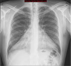
Two signs derive from direct observation of the effusion. The ” pericardial fat stripe ” refers to visualizing fluid density between the epicardial and pericardial fat. As these fat layers are usually more prominent adjacent to the right ventricle, this sign is typically identified anterior to the heart on the lateral radiograph.

What are pericardial fat pads?

Pericardial fat pads are normal adipose tissue masses that lie in the cardiophrenic angles and straddle the pericardium as they are derived from both epicardial fat and epipericardial fat, the latter being adherent and external to the parietal pericardium. Fat pads are more prominent in obese patients.

How do I know if I have a pericardial fat pad?

Epicardial (Pericardial) Fat Pad. Another case: Frontal chest radiograph shows an area of increased density at the left cardiophrenic angle (white arrow) that does not silhouette the heart border. An axial CT scan through the lower thorax shows an accumulation of fat in both the left and right cardiophrenic angles (red arrows).

See more here: What Is The Difference Between Pericardial Effusion And Pericardial Fat Pad? | Fat Pad Sign Pericardial Effusion

Chest Radiograph Signs Suggestive of Pericardial Disease

The “pericardial fat stripe” refers to visualizing fluid density between the epicardial and pericardial fat. As these fat layers are usually more prominent adjacent to the right ventricle, this sign is typically American College of Cardiology

The epicardial fat pad sign: analysis of frontal and lateral chest …

The epicardial fat pad sign (EFPS) has been useful in the diagnosis of pericardial effusion on plain frontal and lateral chest radiographs. In this series of 100 cases, including RSNA Publications Online

Pericardial fat pads | Radiology Reference Article | Radiopaedia.org

Pericardial fat pads are normal adipose tissue masses that lie in the cardiophrenic angles and straddle the pericardium as they are derived from both Radiopaedia

Pericardial effusion and cardiac tamponade – Knowledge – AMBOSS

Pericardial effusion is the acute or chronic accumulation of fluid in the pericardial space (between the parietal and the visceral pericardium) and is often AMBOSS

ECHOCARDIOGRAPHIC ASSESSMENT OF EPICARDIAL FAT

The epicardial fat pad (EFP) is linked to increased risk and the development of major adverse cardiac events. The EFP has also been found to be increased in JACC Journals

Pericardial Effusion Imaging – Medscape

When the pericardial fluid volume is small, it may appear as an anterior hypoechoic or echo-free space behind the left ventricle (LV), which could also represent eMedicine

LearningRadiology – Epicardial, Pericardial Fat, Pad

The epicardial fat pad sign (pericardial fat pad sign, fat pad sign) is an abnormal finding that can be seen with pericardial effusion. Curvilinear fat density in displaced posteriorly from sternum on lateral LearningRadiology

The role of pericardial fat: The good, the bad and the

The pericardial fat volume measured by 64-slice MDCT could be one of the important cardiovascular risk factors, because of the biochemical and biomolecular properties and anatomically close journal-of-cardiology.com

Epicardial Fat: Definition, Measurements and Systematic Review

Epicardial fat (EF) is a visceral fat deposit, located between the heart and the pericardium, which shares many of the pathophysiological properties of other visceral fat deposits, It National Center for Biotechnology Information

See more new information: charoenmotorcycles.com

Epicardial Fat

An Easy Way To Differentiate Between Pericardial Pad Of Fat And Exudative Pericardial Effusion

Pocus Teaching Point Epicardial Fat Pad

Pericardial Fat Pad

What Does Epicardial Fat Look Like In Echo?

Pericardial Or Epicardial Fat Pad Right Ventricle Ultrasound Echocardiography Video

Cardiopulmonary Pocus 6 – Pericardial Effusion

Pericardial Effusion Vs. Fat Pad #Emergencymedicine #Pocus #Echo #Echofirst #Foamed #Foamus

The Pocus Diagnosis Of A Pericardial Effusion: A Practical Guide For Clinicians

Link to this article: fat pad sign pericardial effusion.
Pericardial Effusion | Radiology Reference Article | Radiopaedia.Org
Pericardial Fat Pads | Radiology Reference Article | Radiopaedia.Org
Learningradiology - Epicardial, Pericardial Fat, Pad
Learningradiology – Epicardial, Pericardial Fat, Pad
Untitled Document
Untitled Document
Untitled Document
Untitled Document
Learning Radiology - Pericardial, Effusion, Fluid, Pericardium
Learning Radiology – Pericardial, Effusion, Fluid, Pericardium
Learningradiology - Epicardial, Pericardial Fat, Pad
Learningradiology – Epicardial, Pericardial Fat, Pad
Epicardial Fat Pad | Radiology Case | Radiopaedia.Org
Constrictive Pericarditis And Pericardial Effusion | Radiology Case |  Radiopaedia.Org
Constrictive Pericarditis And Pericardial Effusion | Radiology Case | Radiopaedia.Org
The Epicardial Fat Pad Sign: Analysis Of Frontal And Lateral Chest  Radiographs In Patients With Pericardial Effusion.
The Epicardial Fat Pad Sign: Analysis Of Frontal And Lateral Chest Radiographs In Patients With Pericardial Effusion.
Pericardial Effusion | Ppt
20 Pericardial Effusion | Ppt
Chest Radiology
Chest Radiology
Learning Radiology - Pericardial, Effusion
Learning Radiology – Pericardial, Effusion
Pericardial Effusion
Epos&Trade;
Epos&Trade;
Nephropocus On X:
Nephropocus On X: “#Pocus #Foamed Today’S Lesson: Epicardial Fat Can Mimic Pericardial Effusion. Interestingly, In This Case, Patient Had Tamponade Physiology From Another Cause 😱 Full Case Report (Open Access): Https://T.Co/Xnaq0Xyhvg Https://T.Co …
Pericardial Effusion | Ppt
Pericardial Fatpad Pericardial Fat Depositions Are ... | Grepmed
Pericardial Fatpad Pericardial Fat Depositions Are … | Grepmed
Pocus Teaching Point Epicardial Fat Pad - Youtube
Pocus Teaching Point Epicardial Fat Pad – Youtube
An Easy Way To Differentiate Between Pericardial Pad Of Fat And Exudative Pericardial  Effusion - Youtube
An Easy Way To Differentiate Between Pericardial Pad Of Fat And Exudative Pericardial Effusion – Youtube
Epicardial Fat Pad In Cxr. Posteroanterior Chest Radiography Reveals... |  Download Scientific Diagram
Epicardial Fat Pad In Cxr. Posteroanterior Chest Radiography Reveals… | Download Scientific Diagram
Pericardial Effusion - Radrounds Radiology Network
Pericardial Effusion – Radrounds Radiology Network
Cxr/Tte Before (Left) And After (Right) Pericardial Lipoma Resection.... |  Download Scientific Diagram
Cxr/Tte Before (Left) And After (Right) Pericardial Lipoma Resection…. | Download Scientific Diagram
Pericardial Effusion | Radiology Reference Article | Radiopaedia.Org
Chest Radiograph Signs Suggestive Of Pericardial Disease - American College  Of Cardiology
Chest Radiograph Signs Suggestive Of Pericardial Disease – American College Of Cardiology
Case Studies: Pericardial Effusion And Fat Pad No Disclosures
Pericardial Or Epicardial Fat Pad Right Ventricle Ultrasound  Echocardiography Video - Youtube
Pericardial Or Epicardial Fat Pad Right Ventricle Ultrasound Echocardiography Video – Youtube
Pericardial Effusion And Cardiac Tamponade - Knowledge @ Amboss
Pericardial Effusion And Cardiac Tamponade – Knowledge @ Amboss
Epicardial Fat Sign Due To Extrapericardial Disease
Pericardial Fat Pads | Radiology Reference Article | Radiopaedia.Org
Untitled Document
Untitled Document
Pericardial Fat Tag Sign | Radiology Case | Radiopaedia.Org
Pericardial Effusion: Lateral Radiograph Of The Chest Demonstrating The Pericardial  Fat (Yellow Line) And Ep… | Pericardial Effusion, Medical Radiography,  Radiology
Pericardial Effusion: Lateral Radiograph Of The Chest Demonstrating The Pericardial Fat (Yellow Line) And Ep… | Pericardial Effusion, Medical Radiography, Radiology
Alkhatib Radiology Cases - Oreo Cookie Sign: Lateral Chest Radiograph Shows  A Radiopaque Stripe Separating Two Radio Transparent Stripes, Representing  The Pericardial Effusion Separating The Epicardial And Pericardial Fat Pads,  Which Is
Alkhatib Radiology Cases – Oreo Cookie Sign: Lateral Chest Radiograph Shows A Radiopaque Stripe Separating Two Radio Transparent Stripes, Representing The Pericardial Effusion Separating The Epicardial And Pericardial Fat Pads, Which Is
Pseudo Cardiac Tamponade In The Setting Of Excess Pericardial Fat |  Cardiovascular Ultrasound | Full Text
Pseudo Cardiac Tamponade In The Setting Of Excess Pericardial Fat | Cardiovascular Ultrasound | Full Text
Potential Errors In The Diagnosis Of Pericardial Effusion On Trauma  Ultrasound For Penetrating Injuries
Potential Errors In The Diagnosis Of Pericardial Effusion On Trauma Ultrasound For Penetrating Injuries
The Radiology Assistant : Chest X-Ray - Basic Interpretation
The Radiology Assistant : Chest X-Ray – Basic Interpretation
The Floating Cardiac Fat Pad—Sign Of Occult Pneumothorax | Emergency  Radiology
The Floating Cardiac Fat Pad—Sign Of Occult Pneumothorax | Emergency Radiology
Pericarditis With Pericardial Effusion | Radiology Case | Radiopaedia.Org
What Is Normal? | Kidsono
Pericardial Effusion And Cardiac Tamponade - Knowledge @ Amboss
Pericardial Effusion And Cardiac Tamponade – Knowledge @ Amboss
Pocus Teaching Point Pericardial Effusion - Youtube
Pocus Teaching Point Pericardial Effusion – Youtube
Pericardial Effusion | Radiology Reference Article | Radiopaedia.Org
Pericardial Effusion
Internal Medicine Point Of Care Ultrasound - Impocus
Internal Medicine Point Of Care Ultrasound – Impocus
Pericardial Disease — Tpa
Pericardial Effusion And Cardiac Tamponade - Knowledge @ Amboss
Pericardial Effusion And Cardiac Tamponade – Knowledge @ Amboss
Cardiac Ultrasound (Chapter 17) - Clinical Emergency Radiology
Cardiac Ultrasound (Chapter 17) – Clinical Emergency Radiology
Pericardial Effusion And Cardiac Tamponade - Knowledge @ Amboss
Pericardial Effusion And Cardiac Tamponade – Knowledge @ Amboss
Case Studies: Pericardial Effusion And Fat Pad No Disclosures
Pericardial Effusion | Radiology Reference Article | Radiopaedia.Org
Pericardial Effusion | Radiology Key
Pericardial Effusion Imaging: Practice Essentials, Radiography, Computed  Tomography
Pericardial Effusion Imaging: Practice Essentials, Radiography, Computed Tomography
Learningradiology - Epicardial, Pericardial Fat, Pad
Learningradiology – Epicardial, Pericardial Fat, Pad
Pseudo Cardiac Tamponade In The Setting Of Excess Pericardial Fat |  Cardiovascular Ultrasound | Full Text
Pseudo Cardiac Tamponade In The Setting Of Excess Pericardial Fat | Cardiovascular Ultrasound | Full Text
Pericardial Effusion Imaging: Practice Essentials, Radiography, Computed  Tomography
Pericardial Effusion Imaging: Practice Essentials, Radiography, Computed Tomography
Pericardial Effusion | Radiology Key
The Epicardial Fat Pad Sign: Analysis Of Frontal And Lateral Chest  Radiographs In Patients With Pericardial Effusion.
The Epicardial Fat Pad Sign: Analysis Of Frontal And Lateral Chest Radiographs In Patients With Pericardial Effusion.
Pericardial Fat Pads | Radiology Case | Radiopaedia.Org
Learning Radiology - Pericardial, Effusion
Learning Radiology – Pericardial, Effusion
Epicardial Versus Pericardial Fat Thickness. Pericardial Fat (Within... |  Download Scientific Diagram
Epicardial Versus Pericardial Fat Thickness. Pericardial Fat (Within… | Download Scientific Diagram
Pericardiocentesis – International Emergency Medicine Education Project
Cotw: 3/14/22: 60 Year Old Male With Hoarseness — Kaiser San Diego  Emergency Ultrasound
Cotw: 3/14/22: 60 Year Old Male With Hoarseness — Kaiser San Diego Emergency Ultrasound
Echocardiography In Pericardial Disease | Thoracic Key
Epipericardial Fat Necrosis: An Uncommon Self-Limiting Cause Of Pleuritic  Chest Pain | Cmaj
Epipericardial Fat Necrosis: An Uncommon Self-Limiting Cause Of Pleuritic Chest Pain | Cmaj
Pericardial Effusion | Radiology Reference Article | Radiopaedia.Org
Internal Medicine Point Of Care Ultrasound - Impocus
Internal Medicine Point Of Care Ultrasound – Impocus
Message In A Bottle. The Use Of Chest Radiography For Diagnosis Of Pericardial  Effusion - Sciencedirect
Message In A Bottle. The Use Of Chest Radiography For Diagnosis Of Pericardial Effusion – Sciencedirect
Epos&Trade;
Epos&Trade;
Fat Pad Sign - Print Friendly - Statdx | Pdf | Radiography | Radiology
Fat Pad Sign – Print Friendly – Statdx | Pdf | Radiography | Radiology
Pericardial Disease — Tpa
Heart Attack Acute Chest Pain Injury Stock Vector (Royalty Free) 2149026531  | Shutterstock
Heart Attack Acute Chest Pain Injury Stock Vector (Royalty Free) 2149026531 | Shutterstock
Pericardial Fat Masquerading As Tumor - Pressman - 2010 - Echocardiography  - Wiley Online Library
Pericardial Fat Masquerading As Tumor – Pressman – 2010 – Echocardiography – Wiley Online Library
Pocus 101 On X:
Pocus 101 On X: “A Pericardial Fat Pad Can Be A False Positive For Pericardial Effusion. It Is Usually Located Anteriorly, Echogenic, And Appears To Move With The Heart Contractions. 🔗👉Https://T.Co/Gmoludsvgx Https://T.Co/Sjmhxbeuc0” /
Epipericardial Fat Necrosis: Radiologic Diagnosis And Follow-Up | Ajr
Pericardial Effusion | Radiology Key

See more articles in the same category here: https://charoenmotorcycles.com/how